Wisdom Teeth Removal and Dental X-Rays: Why They Matter
Wisdom teeth have a way of causing trouble quietly. A patient can feel completely fine, show no swelling, report no toothache, and still have a lower third molar pressing into the tooth in front of it or sitting partly trapped under bone. That is one reason dental X-rays matter so much in decisions about Wisdom Teeth Removal. They reveal what the mouth cannot show on its own.
Most people only see a small part of a wisdom tooth, if they see it at all. Dentists and oral surgeons have to think beyond the visible crown. They need to know the angle of eruption, the amount of bone covering the tooth, the root shape, the relationship to nearby teeth, and, in the lower jaw, how close the roots may be to the main sensory nerve. Those details affect whether removal is recommended, how urgent it is, how complex the procedure may be, and what kind of recovery a patient can expect.
The conversation around wisdom teeth often gets simplified into two camps. Either they "have to come out" or "they can stay forever if they don't hurt." Real life is more nuanced. X-rays are what make that nuance possible.
What wisdom teeth actually are
Wisdom teeth are the third molars, usually the last teeth to develop and emerge. They often appear in the late teens to mid-twenties, although development varies from person to person. Some people have all four, some have fewer, and some never develop them at all.
Human jaws do not always have enough room for these final molars to come in cleanly. When there is limited space, the tooth may erupt only partway, stay trapped under the gum, remain fully embedded in bone, or come in at an awkward angle. That angle matters. A wisdom tooth leaning forward into the second molar creates a very different problem than one growing upright with enough room to function.
This is where clinical exams reach their limit. A dentist can inspect the gums, probe around partially erupted teeth, and check for tenderness, decay, or swelling. But none of that shows the full path of eruption under the surface.
Why pain is a poor guide
One of the most common misunderstandings is the belief that no pain means no problem. In dentistry, that is often untrue. Wisdom teeth may damage nearby structures slowly enough that symptoms show up late.
A classic example is the partially erupted lower wisdom tooth. It creates a pocket under the gum flap where bacteria and food debris collect. The patient may notice an occasional bad taste or mild soreness that comes and goes. On an X-ray, though, the tooth may already be angled into the second molar, increasing the risk of decay or bone loss on a tooth that is far more important for chewing.
Another pattern appears when a wisdom tooth remains fully impacted. It may sit silently for years. In some cases, that tooth causes no harm and can simply be monitored. In others, it may contribute to cyst formation, resorption of the neighboring tooth root, or difficult-to-clean areas that compromise long-term oral health. The difference is not guesswork. Imaging helps separate low-risk cases from high-risk ones.
Patients are often surprised to learn that the most urgent reason for removal is not always the wisdom tooth itself. Sometimes the priority is protecting the healthy second molar beside it. Losing or damaging that tooth is a much bigger functional problem than removing a third molar that never erupted properly.
What dental X-rays show that the eye cannot
When planning Wisdom Teeth Removal, X-rays provide a map. That map answers several practical questions.
First, is the tooth present at all? It sounds basic, but not everyone develops all four wisdom teeth. A missing tooth on the X-ray saves everyone from unnecessary concern.
Second, what is the position of the tooth? Wisdom teeth can be vertical, horizontal, mesioangular, distoangular, or positioned in more irregular ways. The angle affects both the likelihood of future trouble and the surgical approach.
Third, how much room is available? Some wisdom teeth are blocked by the jawbone or neighboring teeth. Others have enough space to emerge and function normally.
Fourth, what do the roots look like? Roots can be straight, curved, fused, long, short, or partly formed. A tooth with incomplete root development in a younger patient is often easier to remove than one with fully formed, hooked roots in an older adult.
Fifth, how close is the tooth to critical anatomy? For lower wisdom teeth, the main concern is the inferior alveolar nerve, which runs through the jaw and provides feeling to the lower lip and chin. For upper wisdom teeth, the maxillary sinus may be relevant. X-rays help estimate these relationships before surgery.
That information changes treatment recommendations in very concrete ways. A straightforward erupted tooth may be removed in a general dental office with local anesthetic. A deeply impacted tooth close to the nerve may be better managed by an oral and maxillofacial surgeon, sometimes after further imaging.
The types of X-rays commonly used
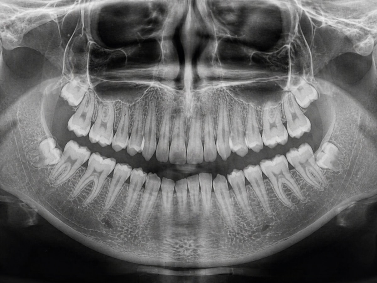
Not every case needs advanced imaging. In many situations, a standard panoramic X-ray gives a broad and useful overview. It shows all the teeth, both jaws, the sinuses, and the general position of the wisdom teeth in one image. For routine screening and initial planning, it is often the workhorse.
A dentist may also use small intraoral X-rays, especially bitewings or periapical films, when checking whether the wisdom tooth has caused decay on the back surface of the second molar. Those smaller views can sometimes reveal detail that a panoramic image does not capture as sharply.
Cone beam CT, often called CBCT, is more specialized. It creates a three-dimensional view and can be extremely helpful when a lower wisdom tooth appears very close to the nerve canal or when anatomy is unusual. It is not necessary for every patient. Good clinicians do not order it automatically. They use it when the added detail is likely to change treatment planning or improve safety.
That point matters because more imaging is not always better. The right image is the one that answers the clinical question. A simple case should stay simple. A complex case deserves a better map.
How X-rays influence the timing of removal
Timing is one of the biggest judgment calls in wisdom tooth care. Remove too early and a patient may go through surgery for a tooth that might never have caused a problem. Wait too long and the procedure can become harder, with denser bone, fully formed roots, and higher complication rates.
X-rays help find the middle ground.
In many younger patients, usually in the mid-teen to early adult years, wisdom teeth are still developing. Roots may be only partly formed, and the bone is generally more forgiving. If the X-ray clearly shows crowding, impaction, or harmful angulation, removal during this stage can be technically easier and recovery may be smoother. That does not mean every teenager needs automatic extraction. It means early imaging helps identify the subset who would benefit from acting before trouble escalates.
For adults in their late twenties, thirties, or beyond, the question shifts. If impacted wisdom teeth have been stable for years and show no evidence of disease, monitoring may be reasonable. But that decision should still be based on radiographic findings, not wishful thinking. A symptom-free tooth with a healthy surrounding structure is different from a symptom-free tooth quietly compromising the molar next to it.
The phrase "watch and wait" sounds passive, but in good dentistry it is active surveillance. That means periodic exams and appropriately timed X-rays, not forgetting the tooth exists.
Cases where X-rays change the plan entirely
A few scenarios come up often in practice.
A patient may arrive expecting all four wisdom teeth to be removed at once. The panoramic X-ray shows that the upper wisdom teeth are erupted, easy to clean, cavity-free, and not causing bite problems, while only the lower teeth are impacted and risky. Suddenly the plan becomes more conservative and more sensible.
In another case, a patient may assume a swollen gum means the wisdom tooth is the only issue. The X-ray reveals decay on the second molar where the wisdom tooth has been trapping plaque. The discussion changes from "take out one tooth" to "remove the wisdom tooth and assess whether the second molar needs a filling, root canal, or more extensive treatment."
Then there is the patient who is worried because a friend had numbness after lower wisdom tooth surgery. On the X-ray, the roots appear well separated from the nerve canal, which supports a lower-risk discussion. Or the opposite happens: the image shows overlap or diversion of the canal, and the surgeon recommends CBCT before deciding on the safest technique. Sometimes that leads to a modified approach, such as coronectomy in select cases, where the crown is removed and the roots are intentionally left to avoid nerve injury. That is not common in every office, but it is a good example of imaging directly affecting judgment.
Why lower wisdom teeth tend to get more attention
Upper wisdom teeth can cause problems, but lower wisdom teeth are usually the more complicated conversation. The lower jaw is denser, access can be tighter, and the inferior alveolar nerve adds an important layer of risk assessment. A lower wisdom tooth may be completely buried in bone, tipped forward under the second molar, or partly erupted in a position that repeatedly traps bacteria under the gum tissue.
This is where patients often see the value of an X-ray immediately. A small ache at the back of the jaw may not seem like much until the image shows a horizontal tooth jammed directly into the molar beside it. On the other hand, a dramatic-looking gum flare-up might come from a tooth that is actually easier to remove than expected because its roots are short and well away from the nerve.
The image does not just detect disease. It gives context.
The risks of skipping imaging
Trying to evaluate wisdom teeth without X-rays is a bit like planning a basement renovation by looking only at the front door. You can gather some useful information, but you are making a major decision without seeing the structures that matter most.
Problems that may be missed without imaging include:
- Decay on the back of the second molar
- Root resorption of the neighboring tooth
- Cystic changes around an impacted tooth
- Unusual root shapes that complicate extraction
- Close nerve proximity that raises surgical risk
Each of these can alter the recommendation, the referral decision, the consent discussion, or the post-operative expectations. They are not minor details.
What patients should ask when reviewing their X-rays
Patients do not need to interpret their own radiographs like a dentist, but they should understand the basics of what the image means for them. A good consultation is not a monologue. It is a conversation grounded in the image on the screen.
Useful questions include whether the tooth is impacted, whether it is harming the tooth next to it, how difficult removal is expected to be, and whether additional imaging is necessary. If the recommendation is to leave a wisdom tooth alone, ask what signs would trigger a change in plan and how often it should be re-evaluated.
This matters because the best decision is not always immediate removal. Sometimes the smartest plan is thoughtful monitoring. Patients deserve to know why one path is being chosen over another.
One detail that often reassures people is seeing the anatomy explained visually. Wisdom Teeth Removal When a surgeon points out the angle of the tooth, the surrounding bone, and the distance from the nerve canal, the decision feels less abstract. Many anxious patients calm down once the problem is made concrete.
Age, anatomy, and recovery
There is a practical reason oral surgeons pay attention to age in wisdom tooth planning. Younger bone tends to be less dense, roots are often not fully formed, and healing is generally efficient. That does not guarantee an easy recovery, but it often improves the odds.
By contrast, removing deeply impacted wisdom teeth in someone older can involve denser bone and more complex root anatomy. Recovery can still go well, but swelling, soreness, and surgical difficulty may increase. This is another place where X-rays sharpen the discussion. They help show whether a thirty-five-year-old with impacted teeth has a stable, low-risk situation that can be monitored, or a ticking problem that has simply not become painful yet.
Dentistry is full of these trade-offs. The right answer depends on the patient in front of you, not a blanket rule.
Dental X-rays and safety concerns
Patients sometimes hesitate because of radiation exposure. That is understandable. Any medical imaging should have a reason behind it. In modern dental practice, the doses from routine dental X-rays are relatively low, especially with digital systems, and the value of the information is often substantial. The key principle is justification. If an image will change diagnosis, planning, or safety, it is serving a clear purpose.
What experienced clinicians try to avoid is both extremes: ordering images casually and avoiding them reflexively. Neither approach serves the patient well. Good care means choosing the minimum imaging needed to make a sound decision.
For wisdom teeth, especially impacted lower third molars, the benefit of seeing the anatomy usually outweighs the downside of remaining blind to it.
When Wisdom Teeth Removal is not the right move
This is an important point because many articles oversimplify it. Not every wisdom tooth belongs in a surgical tray.
A fully erupted wisdom tooth that can be cleaned well, has no decay, has healthy gum support, and does not interfere with the bite may not need removal. Likewise, some fully impacted teeth remain stable for years with no sign of disease. If X-rays support that low-risk picture, observation can be appropriate.
That said, "not removing" is not the same as "never thinking about it again." Circumstances can change. A tooth that looks harmless at age twenty may be associated with a decay problem or gum defect at age twenty-eight. Monitoring intervals depend on the clinical situation, oral hygiene, age, and history of symptoms.
A professional recommendation should feel individualized. If every patient hears the exact same script regardless of anatomy, that Wisdom Teeth Removal is a red flag. X-rays are part of what allows a tailored recommendation rather than a one-size-fits-all one.
What a well-run consultation usually looks like
When wisdom teeth are being evaluated properly, the process is fairly straightforward. The patient describes symptoms, if any. The dentist or oral surgeon examines the mouth and reviews current X-rays. If the anatomy is clear and risk appears routine, treatment can often be discussed right away. If the imaging suggests increased complexity, additional views or referral may follow.
The most helpful consultations usually cover a few practical issues in plain language:
- Whether removal is recommended now, later, or not at all
- How difficult the extraction is expected to be
- What specific risks are relevant in that case
- What recovery is likely to involve
- Whether the X-ray findings affect the choice of provider or sedation
Patients often remember the conversation better when they are shown their own image. It turns a general warning into a personal explanation.
The larger point behind the image
At a glance, a dental X-ray looks simple. Black, white, shades of gray, a few outlines most patients cannot read. But for wisdom teeth, that image often holds the key details that make treatment safer and more precise.
It can prevent unnecessary surgery when a tooth is stable and harmless. It can justify early action when damage is beginning quietly. It can identify when a straightforward extraction is possible and when specialist care is wiser. It can protect nearby teeth, reduce surprises during surgery, and support informed consent that is based on anatomy rather than assumption.
That is why X-rays matter so much in Wisdom Teeth Removal. They do not replace clinical judgment, but they make good judgment possible. Without them, the decision is largely guesswork. With them, the plan can fit the patient, the tooth, and the risk in a way that is measured, practical, and far more likely to hold up over time.
Oxnard Dentistry
1730 E Gonzales Rd, Oxnard, CA 93036, United States
+1 805-307-8731
FAQ About Wisdom Teeth Removal
How painful is it to remove a wisdom tooth?
Local anesthetic numbs the area during extraction. Pressure may still be noticeable; tell the dentist if you feel pain. Soreness and swelling afterward are common. Follow the pain-control instructions provided for your situation.
How long is recovery for wisdom teeth extractions?
Recovery varies with the tooth position and complexity of the extraction. Some people resume routine activities quickly, while others need several days of rest. Soreness, swelling and jaw stiffness can last up to two weeks. Follow your clinician's aftercare and activity advice.
What are the risks of wisdom tooth removal?
Possible complications include dry socket, infection, persistent bleeding and changes in sensation from nearby nerve injury. Your dentist or oral surgeon should explain your individual risks before treatment. Seek prompt dental advice for bleeding that will not stop, fever, or severe or worsening pain and swelling.